
Access more sample types
Compatible with both FFPE and fresh frozen tissue samples.

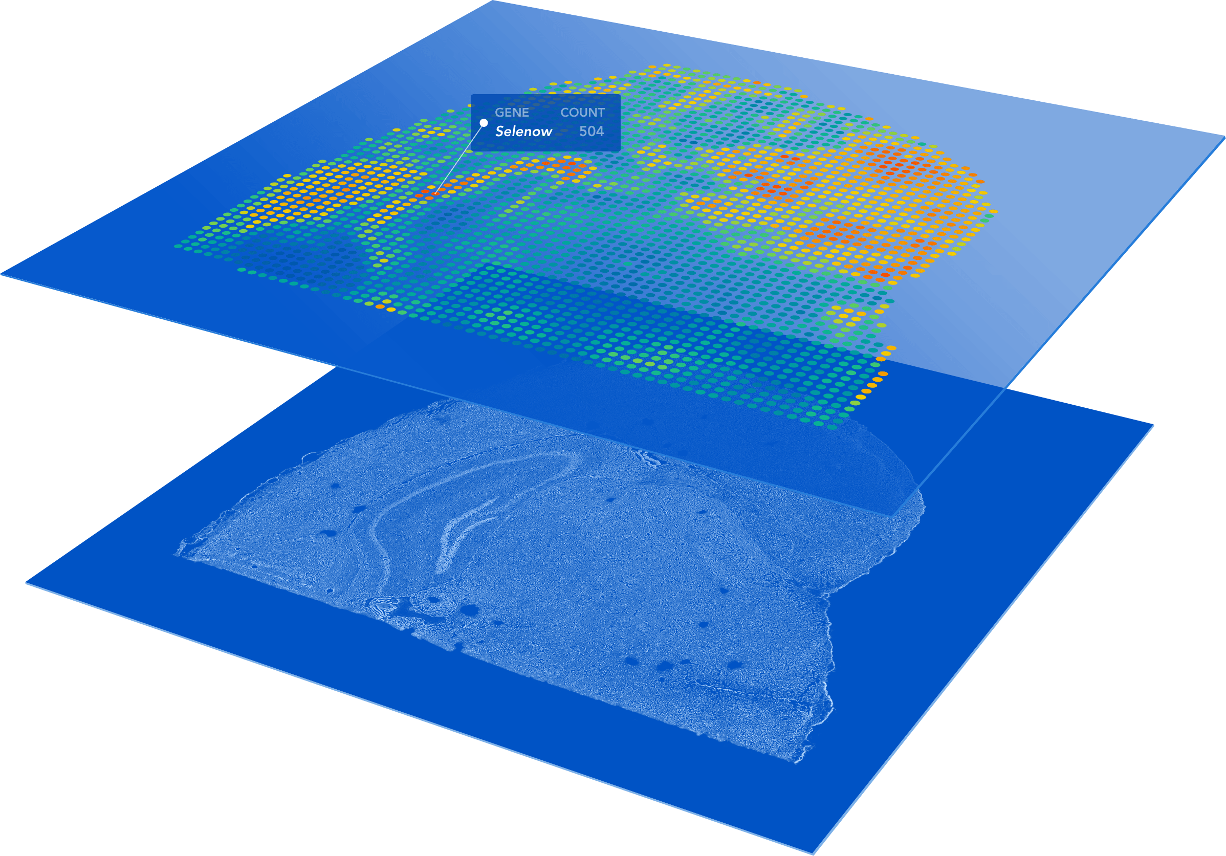
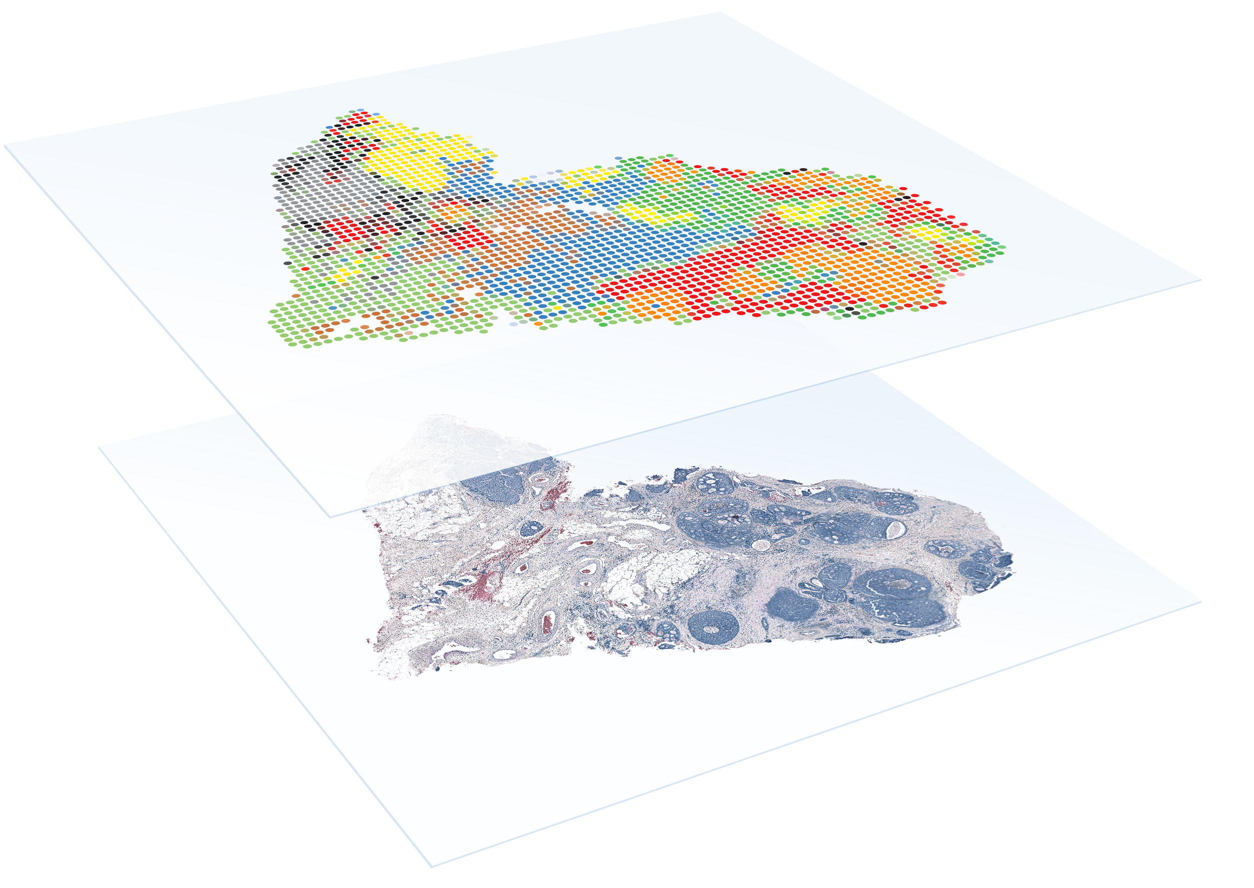
Profile entire tissue sections
No need to select regions of interest—analyze the whole transcriptome from an entire section.

High resolution
1–10 cell resolution on average per spot depending on tissue type.

Diverse sample compatibility
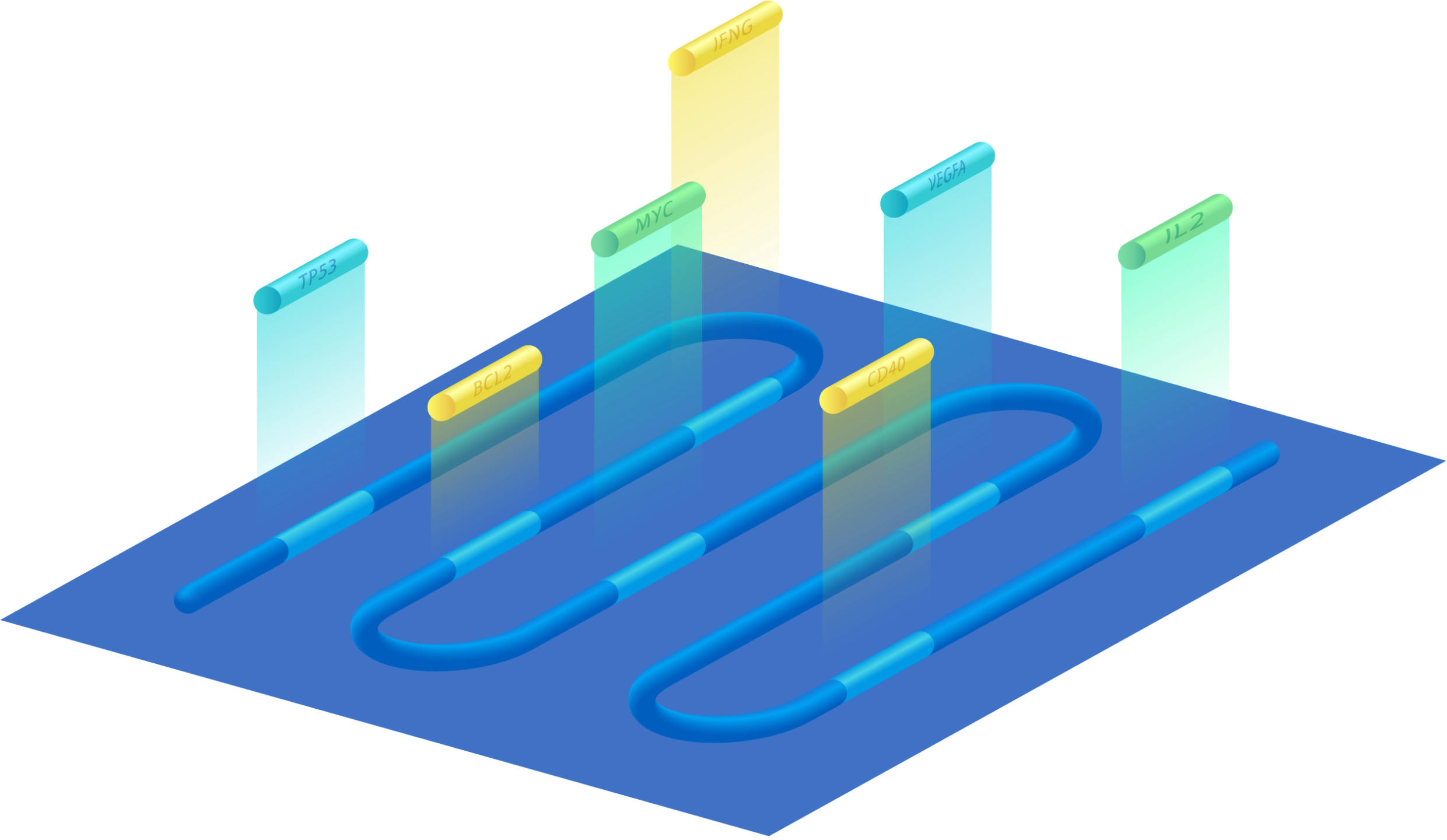
Demonstrated on a diverse set of organs across species (human, mouse, rat, and more).

Protein co-detection
Combine whole transcriptome spatial analysis with immunofluorescence protein detection.

Streamlined data analysis
Combine histological and gene expression data with easy-to-use software.
Explore what you can do
Gain a complete view of disease complexity
Discover new biomarkers
Map the spatial organization of cell atlases
Identify spatiotemporal gene expression patterns
More than a tissue slide
FFPE
FFPE compatibility
Combine the benefits of histological techniques with the massive throughput and discovery power of RNA sequencing to detect the whole transcriptome in FFPE samples.
- Detect the whole transcriptome across entire FFPE tissue sections with high sensitivity and specificity
- Examine biobanked FFPE tissue samples to discover new biomarkers
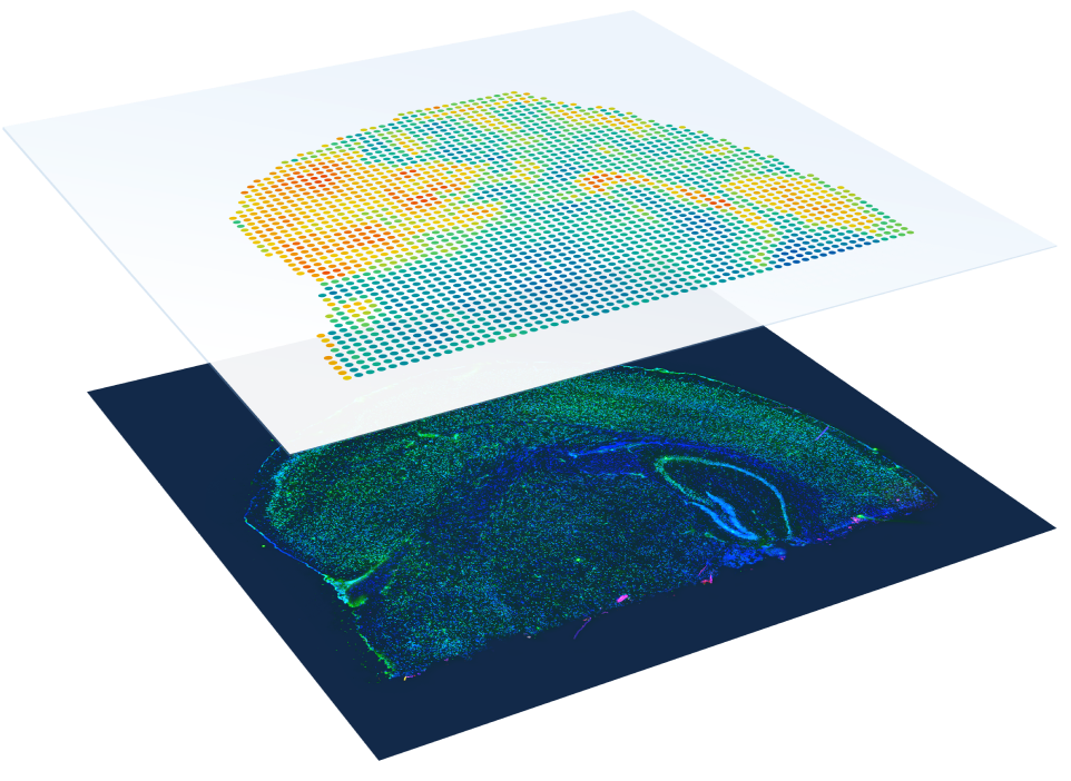
Immunofluorescence
Protein co-detection
Enable discovery of novel cell types and their spatial organization by combining immunofluorescence and whole transcriptome spatial gene expression.
- Explore transcriptome and protein relationships with simultaneous spatial detection
- Discover new tissue biomarkers with cell-type specificity
Flexible transcriptomic analysis
Whole transcriptome or targeted assays
Spatially map global gene expression with whole transcriptome analysis or focus on your genes of interest using targeted gene expression panels.
- Powerful discovery—Envision the spatial organization of newly discovered cell types, states, and biomarkers with whole transcriptome analysis of fresh frozen or FFPE tissues
- Targeted gene expression—Focus on all of the relevant genes and pathways with the convenience of pre-designed targeted panels for fresh frozen tissues
Proven results
Proven results
Publications
Search peer-reviewed publications using Spatial Gene Expression.
Transcriptome-scale spatial gene expression in the human dorsolateral prefrontal cortex
Transcriptome-scale spatial gene expression in the human dorsolateral prefrontal cortex
Maynard KR, et al. Nature Neuroscience 2021.
Maynard KR, et al. Nature Neuroscience 2021.
Spatiotemporal analysis of human intestinal development at single-cell resolution
Spatiotemporal analysis of human intestinal development at single-cell resolution
Fawkner-Corbett D, et al. Cell 2021.
Fawkner-Corbett D, et al. Cell 2021.
TGFβ-blockade uncovers stromal plasticity in tumors by revealing the existence of a subset of interferon-licensed fibroblasts
TGFβ-blockade uncovers stromal plasticity in tumors by revealing the existence of a subset of interferon-licensed fibroblasts
Grauel AL, et al. Nature Communications 2020.
Grauel AL, et al. Nature Communications 2020.
Multimodal analysis of composition and spatial architecture in human squamous cell carcinoma
Multimodal analysis of composition and spatial architecture in human squamous cell carcinoma
Ji AL, et al. Cell 2020.
Ji AL, et al. Cell 2020.
Resources
Find the latest app notes and other Spatial Gene Expression documentation
Inside Visium spatial capture technology
Inside Visium spatial capture technology
Brochure, 10x Genomics
Brochure, 10x Genomics
Grant writing resource for Visium Spatial Gene Expression
Grant writing resource for Visium Spatial Gene Expression
Grant Application Resource, 10x Genomics
Grant Application Resource, 10x Genomics
Triple negative breast cancer app note
Triple negative breast cancer app note
App Note, 10x Genomics
App Note, 10x Genomics
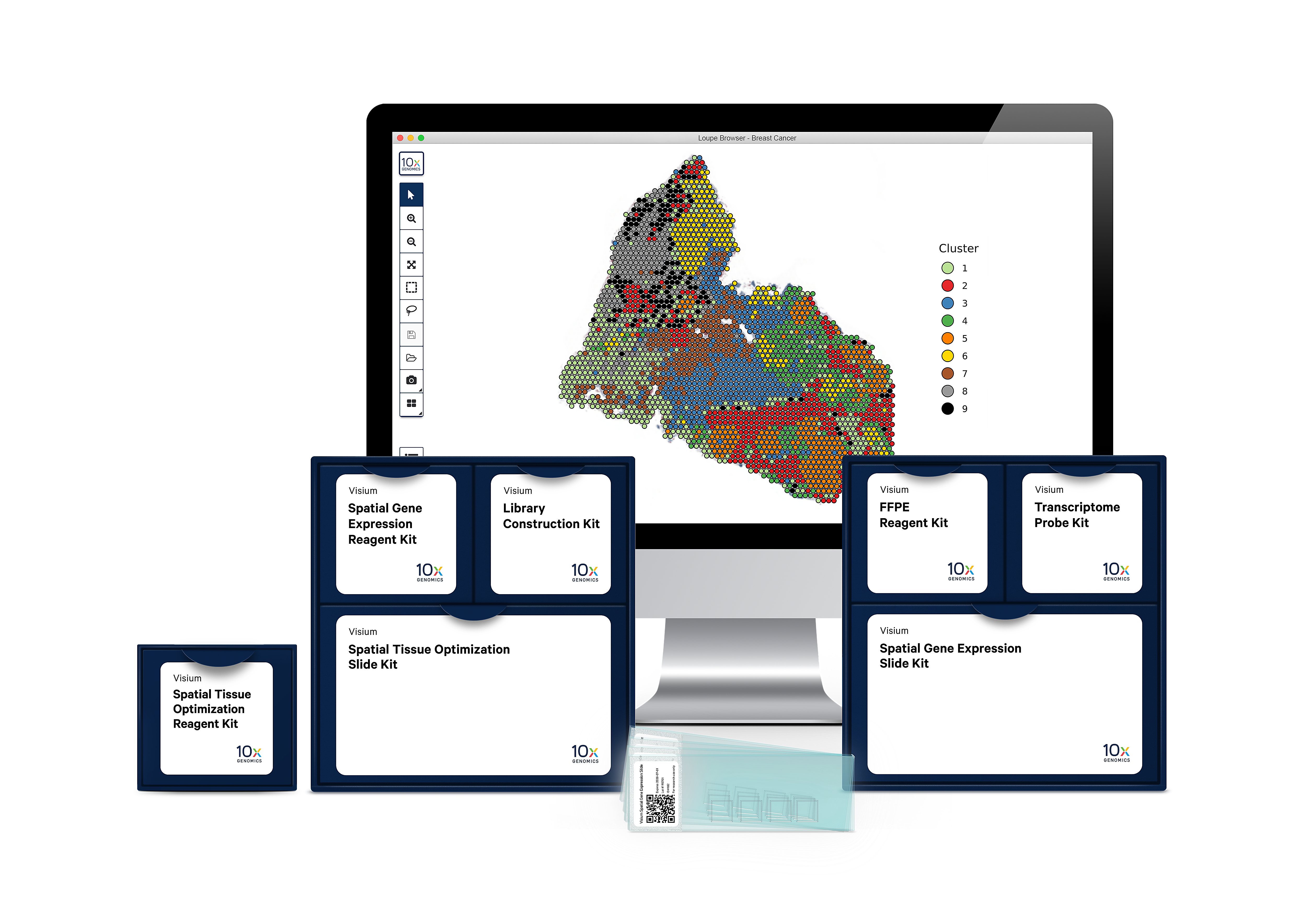
Our end-to-end solution

Visium Spatial Gene Expression Slides
Easily adoptable within existing lab infrastructure, no need for a new instrument.

Visium Spatial Gene Expression Reagents
With our reagent kits, capture whole transcriptome gene expression with spatial resolution across an entire tissue section.

Analysis and visualization software
Our analysis pipelines
Our visualization software

World-class technical and customer support
Our expert support team can be contacted by phone or email.
Workflow
- 1

Prepare your sample
Embed, section, and place fresh frozen or FFPE tissue onto a Capture Area of the gene expression slide. Each Capture Area has thousands of barcoded spots containing millions of capture oligonucleotides with spatial barcodes unique to that spot.
Resources- Tissue Preparation Guide
- Optimized Fresh Frozen Tissues
- Fresh Frozen How-To Videos
FFPE Tissues Tested (Coming Soon)
FFPE How-To Videos (Coming Soon)
- 2

Stain and image the tissue
Utilize standard fixation and staining techniques, including hematoxylin and eosin (H&E) staining, to visualize tissue sections on slides using a brightfield microscope and immunofluorescence staining to visualize protein detection in tissue sections on slides using a fluorescent microscope.
Resources- Imaging Guidelines
- Visium Fresh Frozen User Guide
Visium FFPE User Guide (Coming Soon)
- 3

Permeabilize tissue and construct library
For fresh frozen tissue, the tissue is permeabilized to release mRNA from the cells, which binds to the spatially barcoded oligonucleotides present on the spots. A reverse transcription reaction produces cDNA from the captured mRNA. The barcoded cDNA is then pooled for downstream processing to generate a sequencing-ready library. For FFPE tissues, tissue is permeabilized to release ligated probe pairs from the cells, which bind to the spatially barcoded oligonucleotides present on the spots. Spatial barcodes are added via an extension reaction. The barcoded molecules are then pooled for downstream processing to generate a sequencing-ready library.
Resources- Visium Fresh Frozen User Guide
Visium FFPE User Guide (Coming Soon)
- 4

Sequence
The resulting 10x barcoded library is compatible with standard NGS short-read sequencing on Illumina sequencers for massive transcriptional profiling of entire tissue sections.
Resources - 5

Analyze and visualize your data
Use our Space Ranger analysis software to process your spatial gene expression data and interactively explore the results with our Loupe Browser visualization software.
Do I need to be a bioinformatician to use Loupe?
Loupe is a point-and-click software that’s easy for anyone to download and use.
Resources
Frequently Asked Questions
It enables you to perform spatial transcriptomics, which combines histological spatial information with whole transcriptome gene expression profiles (fresh frozen or FFPE) or targeted gene expression profiles (fresh frozen only).
Visium is compatible with fresh frozen and FFPE tissues containing mRNA. We have separate kits that utilize different chemistries for fresh frozen and FFPE tissues. A list of tissues successfully tested in-house with the Visium assay can be found below.
Successfully tested tissues: Fresh Frozen | FFPE tissues
Visium is based on Spatial Transcriptomics technology, which has been demonstrated by a number of published peer review articles. You can find them on our publications page. For external validation of Visium, beta testers (including Sanger, SciLife, Broad, USC, and Garvan) have been able to generate spatial gene expression profiles for a number of different sample types. Internally, over 30 different fresh frozen tissues and a variety of different FFPE tissues have been tested successfully.
10x Genomics provides two types of software to help you analyze your data: Space Ranger and Loupe Browser. Space Ranger is an analysis software which automatically overlays spatial gene expression information on your tissue image and identifies clusters of spots with similar transcription profiles. You can then use Loupe Browser, a visualization software, to interactively explore the results. You can download Space Ranger and Loupe Browser from the 10x Support site at no cost.
The Visium Spatial Gene Expression assay for FFPE tissues does not require tissue optimization. However, the Visium Spatial Gene Expression assay for fresh frozen tissues does require tissue optimization due to differences in the chemistry and molecular biology between the Visium assays for the two sample preparation types.